


Абстракт
В статье представлены данные об изменениях маркеров нарушения метаболизма в печени (аланинаминотрансфераза — АЛТ, аспартатаминотрансфераза — АСТ, гамма-глутамилтрансфераза — ГГТ, лактатдегидрогеназа — ЛДГ) в крови и лимфе, о скорости лимфотока (СЛТ), а также о гистоморфологических особенностях печени при аллоксан-индуцированном сахарном диабете у кроликов. Экспериментальные исследования были проведены на 14 кроликах обоего пола. Животные были разделены на 2 группы: контрольную (6 кроликов) и экспериментальную (8 кроликов). Диабетические симптомы были выявлены на 5-й день после инъекции аллоксана. Для анализа брали кровь из полости сердца, а лимфу — из грудного лимфатического протока. Операции проводились под анестезией. У кроликов обеих групп исследовали уровень глюкозы, маркеры нарушения метаболизма печени, скорость лимфотока, а также гистоморфологические особенности печени. У животных экспериментальной группы показатели определялись на 5-й, 15-й и 30-й дни после введения аллоксана. Установлено, что повышение маркеров нарушения метаболизма печени как в крови, так и в лимфе, а также развивающиеся гистоморфологические изменения в печени сопровождались снижением скорости лимфотока
Сопроводительное письмо
Diabetes mellitus (DM) is a rapidly spreading disease worldwide, characterized by long-term complications, and remains one of the serious medical and social problems. According to the World Health Organization, in 2021, the number of people with diabetes worldwide exceeded 500 million, and it is projected that this indicator will reach 700 million by 2030 [ 1, 2, 3]. Diabetes is also spreading rapidly in Azerbaijan. In 2024, the total number of registered diabetic patients in the unified electronic registry of medical institutions under the Administration of the Regional Medical Divisions was reported to be 322,353 [4, 5 ].
One of the organs most affected by DM is the liver. The liver plays a key role in regulating glucose homeostasis. During persistent hyperglycemia, end-products of non-enzymatic glycosylation accumulate in the body, including hepatocytes. This leads to damage of both intracellular and extracellular structures, a decrease in NADPH levels, an acceleration of oxidative stress, and increased conversion of glucose to sorbitol [6, 7, 8, 9, 10, 11]. As a result of the disruption of the integrity of hepatocyte membranes, enzymes such as ALT, AST, GGT, and LDH are released into the blood and lymph. This is considered an indicator of liver damage. Although significant progress has been made in studying the histomorphological changes in the liver and the alterations in the levels of enzymes considered indicators of liver damage in the blood in diabetes mellitus, however, there is limited information about such changes in the lymph under conditions of persistent hyperglycemia [12, 13, 14, 15]. It should be noted that a significant portion of the lymph in the thoracic duct originates from the liver, and lymphatic fluid can provide more detailed information about metabolic processes occurring in tissues [6, 17, 18]..Although the role of the lymphatic system in the pathogenesis of liver failure is of great importance, its role during diabetes mellitus has not been sufficiently investigated [19, 20, 21,22].
The aim of this research was to comparatively study the levels of markers impaired metabolism in the liver (ALT, AST, GGT, LDH) in the blood and lymph during experimentally induced diabetes mellitus in rabbits using alloxan injection, to determine the lymph flow rate, and to investigate histomorphological changes in the liver.
MATERIALS AND METHODS The study was carried out on 14 rabbits of the “Chinchilla” breed, weighing between 1.8 and 2.3 kg. The rabbits used in the experiments were divided into two groups: the control group included 6 animals, and the experimental group included 8. In the experimental group, diabetes mellitus was induced in rabbits by intraperitoneal administration of 5% alloxan monohydrate solution at a dose of 100 mg/kg. Diabetic symptoms were observed 5 days after the injection. Blood samples were obtained from the cardiac cavity and lymph samples from the thoracic duct. All procedures were carried out under anesthesia. For this purpose, 8 mg/kg of Calypsol and 0.15 ml/kg of 1% Dimedrol solution were injected intravenously. In the control group, blood and lymph samples were collected from six rabbits, and the levels of liver function–related enzymes (ALT, AST, γ-GT, LDH) as well as LFR were determined. In addition, the histomorphological structures of the liver were studied. The same parameters were assessed in the experimental group of 8 rabbits on 5th, 15th, and 30th days after alloxan administration. For histomorphological evaluation, liver tissues were fixed using 10% neutral buffered formalin or Bouin’s solution. Subsequently, the specimens were dehydrated through a graded ethanol series, embedded in paraffin, sectioned, and stained for histological examination. The numerical data obtained during the course of the study were statistically analyzed in accordance with current recommendations. Statistical analyses were carried out using the method of variation analysis and with the help of the ‘IBM Statistics SPSS-26’ software. For the comparison of variation series, the non-parametric Mann-Whitney U test was also used.
RESULTS AND DISCUSSION During the study, the levels of enzymes characterized the diabetic hepatopathy (ALT, AST, γ-GT, LDH), LFR, and the histomorphological structures of the liver were investigated in the blood and lymph of rabbits with alloxan-induced diabetes mellitus over a period of 30 days and compared with control values. The obtained results are presented in the tables below (Table 1).
As seen from Table 1, after alloxan administration, the levels of blood glucose and the liver function-assessing enzymes (ALT, AST, GGT, LDH) gradually increased throughout the experiment (p<0,001). The dynamics of changes in the parameters determined in the lymph are in the same direction as in the blood (Table 2). As seen from Table 2, after alloxan administration, the levels of glucose and liver function-assessing enzymes (ALT, AST, GGT, LDH) in the lymph gradually increased (p<0.001), and the lymph flow rate decreased throughout the experiment (Table 2).
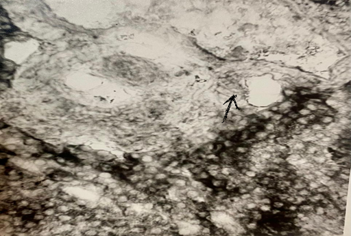
As seen from the presented tables, in animals with induced diabetes mellitus, an increase in the activity of ALT, AST, GGT, and LDH in blood and lymph was observed throughout the experiment compared to the control values. This tendency was more pronounced in the lymph than in the blood. In rabbits with alloxan-induced diabetes mellitus, the increased markers of metabolic disorders in the liver (ALT, AST, GGT, LDH) in blood and lymph are explained by the acceleration of hepatocyte cytolysis under persistent hyperglycemia [23, 24, 25]. During the experiment, histomorphological changes developing in the liver were studied on the 5th, 15th, and 30th days after alloxan administration. On the 5th day of alloxan-induced diabetes mellitus, dystrophic changes were observed in hepatocytes around the periportal vessels (Figure 1). Based on the information presented above, we conclude that during diabetes mellitus, enzymes released from damaged hepatocytes enter the bloodstream and lymphatic system. The increase of these enzymes in both blood and lymph, along with the histomorphological changes occurring in the liver, is considered the development of diabetic hepatopathy, one of the long-term complications of diabetes mellitus [26, 27, 28, 29 ]. The decrease in lymph flow rate slows the removal of accumulated toxic products from tissues, further deepening the histomorphological changes that are developing in the liver [30, 31,32,33].
CONCLUSİON. In alloxan-induced diabetes mellitus, the increased markers of metabolic disorders in the liver (LDH, AST, ALT, GGT) in both blood and lymph, along with the progressive deepening of histomorphological changes in the liver, were accompanied by a decrease in lymph flow rate.
Фигуры
Ключевые слова
Ссылки
1.Sun H., Saeedi P., Karuranga S., et.al. IDF Diabetes Atlas: Global, regional and country level diabetes prevalence estimates for 2021 and projections for 2045., Diabetes Res Clin Prac. 2022, Jan:183:109119. doi: 10.1016/j. diabres. 2021. 109 119. Epub 2021 Dec 6.,https://pubmed.ncbi.nlm.nih.gov/34879977/
2.Paschou S. A., Papadopoulou-Marketou N. et.al. On Type 1 Diabetes Mellitus Pathogenesis, Endocr Connect 2018, Jan, 7(1), p.38-46. PMID: 29191919, PMCID: PMC5776665, DOI: 10.1530/EC-17-0347, https://pubmed.ncbi.nlm.nih. gov/29191919/
3.Abu N. A., Lim C. B., Nor N. S. M. Glycogenic Hepatopathy in Children with Poorly Controlled Type 1 Diabetes Mellitus, Clin Pediatr Endocrino, 2021, 30(2), p.93-97. PMID: 33867669,PMCID: PMC8022034, DOI: 10.1297/cpe.30.93,https://pubmed.ncbi.nlm.nih.gov/33867669/
4.Abdulla A. Number of diabetics in Azerbaijan announced, Administration of the Regional Medical Divisions, 2024
5.Alieva T.T. Changes in prevalence of diabetes mellitus in the Republic of Azerbaijan in 1994-2012 and prospects for 2030, Kazan Medical Journal, Home Archives 2014, Vol 95, No 4. DOI: https:// doi.org/ 10. 17816/KMJ1845, https://kazanmedjournal.ru/kazanmedj/article/view/1845?utm_source=chatgpt.com
6.Berzigotti A., Tsochatzis E., Boursier J., et.al. EASL Clinical Practice Guidelines on Non-invasive Tests for Evaluation of Liver Disease Severity and Prognosis, Update. J. Hepatol, 2021, Volume 75, Issue 3, September p. 659-689. https://www.scirp.org/reference/referencespapers?referenceid=4041224
7.Jessica E B F Lima , Natalia C S Moreira, Elza T Sakamoto-Hojo Mechanisms underlying the pathophysiology of type 2 diabetes: from risk factors to oxidative stress, metabolic dysfunction, and hyperglycemia, Mutat Res Genet Toxicol Environ Mutagen, 2022, Feb-Mar:874-875:503437. PMID: 35151421 DOI: 10.1016/j.mrgentox.2021.503437 https://pubmed.ncbi.nlm.nih.gov/35151421/
8.Jiang S., Tang X., Wang K. et.al. Hepatic functional and pathological changes of type 1 diabetic mice in growing and maturation time, J Cell Mol Med., 2019, 23(8), p. 5794–5807. DOI: 10.1111/ jcmm. 14504, https://doi.org/10.1111/jcmm.14504, https://pubmed.ncbi.nlm.nih.gov/31222979/
9.Najafova V.N. Non- invasive radiodiagnosis of hepatic fibrosis in diabetic patients, Azerbaijan Tibb Jurnalı, Bakı, 2024, №2, p.97-103. DOI: https:doi.org/10.34921/amj.2024.80.94.002, http://azmedjournal.com/index.php/amz/article/view/461
10.Dadashova A., Aliyeva G., et.al. Hyperhomocysteinemia in pediatric β- thalassemia: links to vitamin cofactor deficiencies and oxidative stress, Clin Exp Pediatr, 2025, Jul 8,PMİD:40776617. https://pubmed.ncbi.nlm.nih.gov/40776617/
11.Rahimova R.R. Study of matrix metalloproteinase activity in patients with autoimmune thyroiditis, Ukr.Biochem.J, 2022, Volume 94. İssue, p. 51-56. http://ukrbiochemjournal.org/2022/07/study-of-matrix-metalloproteinase-activity-in-patients-with-autoimmune-thyroiditis.html
12.de Vries M., Westerink J., Kaasjager K.H.A.H., de Valk H.W. Prevalence of nonalcoholic fatty liver disease (NAFLD) in patients with type 1 diabetes mellitus: a systematic review and meta-analysis, J Clin Endocrinol Metab.,2020, 105(12), p. 3842–3853. DOI: 10.1210/clinem/ dga 575?https://pubmed.ncbi.nlm.nih.gov/32827432/
13.Shiful I., Sadaqur R., et.al. Prevalence of elevated liver enzymes and its association with type 2 diabetes: A cross-sectional study in Bangladeshi adults, Endocrinol Diabetes Metab, 2020, Feb 12;3(2):e00116. doi: 10.1002/edm2.116, PMCID: PMC7170449 PMID: 32318634https://pmc.ncbi.nlm.nih.gov/articles/PMC7170449/#:~:text=In%20this%20study%2C%206 1.2%25%20of,higher%20in%20subjects%20having%20diabetes
14.Rafaqat S., Sattar A., Khalid A. et.al. Role of liver parameters in diabetes mellitus – a narrative review, Punjab, Endocrine regulations, 2023, vol. 57, 200–220, doi:10.2478/enr-2023-0024, https://www.lvrach.ru/2021/12/15438168?utm_source=chatgpt.com, https://www.sav.sk/journals/uploads/02261650EndoReg_2023_57_08_Rafaqat.pdf
15.Bueverov A.O., Zilov A.B. Liver damage in type 1 diabetes mellitus, Russian Journal of Gastroenterology, Hepatology, Coloproctology, 2021, Том 31, № 2.https://doi.org/10.22416/1382-4376-2021-31-2-7-13.
16.Mandal A., Bhattarai B., Kafle P. et.al. Elevated Liver Enzymes in Patients with Type 2 Diabetes Mellitus and Non-alcoholic Fatty Liver Disease, National Library of Medicine, 2018, Nov 23, 10(11): p. 3626. PMCID: PMC 6347442 PMID:30697502, https://pmc.ncbi.nlm.nih.gov/articles/PMC6347442/, https://pubmed.ncbi.nlm.nih.gov/30697502/
17.Asada S., Kawaratani H., Mashitani T. et.al. Glycogenic hepatopathy in type 1 diabetes mellitus,2018, Intern Med 57, 1087–1092. PMID: 29279489, PMCID: PMC5938497, DOI: 10.2169/internalmedicine.9490-17, https://pubmed.ncbi.nlm.nih.gov/29279489/
18.Rzayev Z.I, Аkhundov I.T., .Ibrahimli Sh. F., Islamzade F.I. The influence of endolymphatic drug therapy on the functional indexes in patients with lower extremities diabetic angiopathy and diabetic foot syndrome. Azerbaijan Tibb Jurnalı, Bakı,2024, №1, 96-102, DOI: 10.34921/amj.2024.1.015.
19.Abboud W., Abdulla S., Al Zaabi M., Moufarrej R. Young Man with Hepatomegaly: A Case of Glycogenic Hepatopathy, Case Reports Hepatol, 2018, Apr 15:6037530. PMID: 29850300, PMCID: PMC5925151,DOI: 10.1155/2018/6037530,https://pubmed.ncbi.nlm.nih.gov/29850300/
20.Noroozi Karimabad M, Khalili P., Ayoobi F. et.al. Serum liver enzymes and diabetes from the Rafsanjan cohort study, BMC Endocr. Disord, 2022, 12, 22(1), 127. doi: 10.1186/s12902-022-01042-2.,PMID: 35549705,PMCID: PMC9102258, DOI: 10.1186/s12902-022-01042-2, https://pubmed.ncbi.nlm.nih.gov/35549705/
21.Yaru Bi , Yang Yang , Xiaojie Yuan , et.al. Association between liver enzymes and type 2 diabetes: a real-world study, Front Endocrinol (Lausanne), 2024, Feb 20, 15, 1340604. PMCID: PMC 1091 3017PMID:384 44589, https://pmc.ncbi.nlm.nih.gov/articles/PMC10913017/, https://pubmed.ncbi.nlm.nih.gov/38444589/
22.Delli Bovi A.P., Marciano F. et.al. Oxidative Stress in Non-alcoholic Fatty Liver Disease, An Updated Mini Review. Review article, Front. Med.,26 February, Sec. Gastroenterology, 2021, Vol.8. https://www.frontiersin.org/journals/medicine/articles/ 10.3389/fmed.2021.595371/full?
23.Li Y, Wang J, Han X, Hu H. et.al. Serum alanine transaminase levels predict type 2 diabetes risk among a middle-aged and elderly, Chinese population. Ann Hepatol 2019, 18, p. 298–303. https://pubmed.ncbi.nlm.nih.gov/31040092/
24.Wu X, Xu J. Relationship between aspartate aminotransferase to alanine aminotransferase ratio and risk of incident diabetes mellitus in a Chinese population, Second Affiliated Hospital & Yuying Children's Hospital of Wenzhou Medical University, Research Square. 2022, DOI: https://doi.org/10.21203/rs.3.rs-1369408/v1
25.Wang Y, Wu T, et.al. Relationship between serum gamma-glutamyl transferase level and impaired fasting glucose among, Chinese community-dwelling adults: a follow-up observation of 6 years. Metab Syndr Relat Disord 19, 2021, 100–106. https://pubmed.ncbi.nlm.nih.gov/33170087/
26.Kong M, Liu C et.al. Higher level of GGT during mid-pregnancy is associated with increased risk of gestational diabetes mellitus, Clin Endocrinol 2018, 88, p.700–705. https://pubmed.ncbi.nlm.nih.gov/29385633/
27.Kashtl GJ, Abed BA et.al. A comparative study to determine LDH enzyme levels in serum samples of women with breast cancer and women with breast cancer and type 2 diabetes mellitus, J Med Chem Sci 2023, 6, 883–890
28.Talishinskaya M.B., İbrahimova G.H., Panahova S.R., et.al. Diabetic hepatopathy as a manifestation of the late complications of diabetes mellitus, ATU Journal (2025), Volume 5 №2, p.23-33, DOİ: https://doi.org/10.28942/atuj.v5i2y2025.131
29.Talishinskaya M.B., Ibrahimova G.H., Mehdiyev T.V. Correction of markers of diabetic
osteopathy in blood and lymph, АТJ, 2025, №2, 177-181. DOI: https//doi.org/10.34921/amj.2025.2.028
30.Ahmed F. W., Kirupakaran T., Quante M., Smith A. Hepatomegaly, Elevated Hepatic Enzymes, and Bridging Fibrosis in Patients with Type 1 Diabetes Mellitus, Cureus, 2021, Apr 13, 13(4), e14446. PMID: 33996309, PMCID: PMC8114958, DOI: 10.7759/cureus.14446, https://pubmed.ncbi.nlm.nih.gov/33996309/
31.Dmour HH, Khreisat EF, et.al. Assessment of lactate dehydrogenase levels among diabetic patients treated in the outpatient clinics at King Hussein Medical Center, Royal Medical Services, Jordan. Med Arch, 2020, 74, 384–386. https://pubmed.ncbi.nlm.nih.gov/33424095/
32.Bancroft JD, Gamble M. Bancroft's Theory and Practice of Histological Techniques. 8th ed. Elsevier; 2019.
33.Suvarna SK, Layton C, Bancroft JD. Bancroft's Theory and Practice of Histological Techniques. 9th ed. Elsevier; 2024.
О статье:

История публикации

Опубликовано: 13.Oct.2026

Авторское право
© 2022-2025. Azerbaijan Medical University. E-Journal is published by "Uptodate in Medicine" health sciences publishing. All rights reserved.Связанные статьи
ВЛИЯНИЕ ПАТОЛОГИЙ ЩИТОВИДНОЙ ЖЕЛЕЗЫ НА ДИАБЕТОГЕННЫЕ МЕХАНИЗМЫ ПРИ САХАРНОМ ДИАБЕТЕ
Просмотрено: 12