


Абстракт
В настоящем исследовании изучены морфометрические характеристики сосудов толстого кишечника у человека в нормальных условиях, а также в экспериментальных моделях на животных. С использованием микроскопических методов измерялись диаметры, длина, плотность и площадь поперечного сечения артериол, прекапилляров, капилляров, посткапилляров и венул. Нормативные данные выявили региональные различия в распределении сосудов в пределах толстого кишечника, тогда как экспериментальные результаты продемонстрировали морфологические изменения сосудов при кровотечении и нарушениях перфузии. Полученные данные предоставляют ценные сведения о функциональном состоянии сосудистой системы толстого кишечника, расширяют понимание ишемического и постишемического патогенеза и способствуют совершенствованию диагностических и терапевтических стратегий в клинической гастроэнтерологии
Сопроводительное письмо
INTRODUCTION The digestive process consists of sequential stages characterized by the formation of the food bolus, the production of chyle, and the absorption of substances into the blood and lymph. This system plays a crucial role in maintaining homeostasis in the organism, and the biological meaning of its functions is related to the formation of blood plasma. Since the nutrients entering the blood and lymph are foreign to the organism, each food intake represents one of the main challenges to homeostasis [1].
In modern times, comprehensive investigation of the vascular bed of the digestive system organs remains an urgent problem. Only precise information about the state of blood flow can provide an understanding of the morphological mechanisms regulating intramural circulation [2,3]. According to literature data, the gastrointestinal organs receive a large portion of the cardiac output, thereby contributing to the maintenance of metabolism. This process is evidenced by vascular reactions occurring during increased nutrient absorption. The blood circulation of the gastrointestinal organs also supports the protective mechanisms of the mucosa after the intake of toxins or harmful substances and is necessary to ensure adequate perfusion of vital organs during stress. Under certain physiological (exercise) and pathological (hemorrhage) conditions, a sharp decrease in blood flow in the gastrointestinal organs is observed [2].
In recent years, advances in angiological research, microsurgery, endoscopy, clinical lymphology, and gastroenterology have enabled more detailed studies of colonic vascularization. The investigation of the morphological features of colonic blood vessels is important not only from a theoretical perspective but also for improving diagnostic and therapeutic strategies in clinical gastroenterology.
Aim: The aim of this study is to analyze the morphometric parameters of colonic blood vessels in humans under normal conditions and in experimental animals, to determine their structural organization and functional state, and to evaluate the potential applications of these findings in improving diagnostic and therapeutic strategies in clinical gastroenterology.
MATERIALS AND METHODS The study analyzed preparations obtained from cadavers of both sexes aged 20–70 years, in which vascular occlusions and stenoses had been identified. Experimental investigations were conducted on 12 dogs of both sexes, weighing between 10.0 and 18.0 kg (9 animals in the main group and 3 in the control group). The housing and euthanasia of animals were performed in accordance with Directive 2010/63/EU of the European Parliament and the Council on the protection of animals used for scientific purposes [4].
To achieve the stated objectives, the experimental part of the study employed a universal method based on classical impregnation techniques (intravascular Ranvier–Coyer and immersion Bilshovsky–Gross methods). This method was developed by I.I. Markov (Certificate of Authorship No. 1919100, dated September 8, 1990) and had been previously tested in studies of microvascular blood flow in laboratory animals [5,6]. Histological sections were examined using microscopic techniques. Prior to staining, paraffin sections were placed in 96% ethanol for 20–30 minutes to remove residual fat. The deparaffinized sections were rinsed in distilled water, stained with Ehrlich’s hematoxylin solution for 2–5 minutes, washed again in distilled water, and subsequently in running tap water for 3–5 minutes. Differentiation was performed for 1–2 seconds in 1% hydrochloric acid solution prepared in 70° ethanol. Sections were then washed in running water for 30 minutes with frequent changes. The prepared slides were mounted with cover glasses and examined under a light microscope. For Van Gieson staining, Weigert’s iron hematoxylin solution and a mixture of picric acid with fuchsin were used. Pre-prepared sections were immersed in Weigert’s hematoxylin for 2–5 minutes, rinsed in distilled water for 10 minutes, and subsequently placed in the picrofuchsin solution for 2–3 minutes. The sections were cleared in a carbol–toluol solution and mounted in Canada balsam for microscopic evaluation.
RESULTS AND DISCUSSION New data have been obtained regarding the histostructures of the longitudinal layer of the muscular coat. It was revealed that this layer is not continuous along the circumference of the intestine but consists of separate segments (Fig.1). In preparations, the number of these segments varied from 8 to 16, with the distance between individual segments ranging from 25 to 525 μm, and their length from 300 to 1200 μm. The intersegmental gaps along the intestinal circumference were recorded to be arranged in a clockwise direction, indicating that the longitudinal layer of the muscular coat moves in a spiral manner. At the mesenteric border of the intestine, the intersegmental gaps of the longitudinal layer are constant and always coincide with portions of the circular layer of the muscular coat. In this way, muscular “gates,” or rather muscular “tunnels,” are formed, through which straight arteries enter the submucosal base, while straight veins are directed toward the mesentery. The straight vessels pass through the muscular coat at angles ranging from 65° to 850°. Under physiological conditions, the area of intersegmental gaps along the intestinal perimeter constitutes between 1.98% and 4.77% of the ring area. This means that during intestinal peristalsis, the conductivity of the vascular bed may both increase and decrease by up to 2.5 times.
Arteries passing through the muscular “gates” divide into two branches of equal diameter, forming the main submucosal vascular plexus of the intestine. In cross-sections, the relationship of the longitudinal layer of the muscular coat with arteries, arterioles, veins, venules, lymphatic vessels, and capillaries traversing it is clearly visible (Fig. 2). The functional range of these vessels (from complete compression between muscular segments to maximal dilation) demonstrates the influence of the longitudinal muscular layer on the vascular lumen. Notably, there is no loose connective tissue between the walls of lymphatic vessels and capillaries and the muscular segments.
Thus, the results of studying the vasculature of the human large intestine under normal conditions show that the extra-organ arteries supplying blood to this organ (celiac trunk, superior and inferior mesenteric arteries) are of the muscular-elastic type. Their walls exhibit clearly defined external and internal elastic membranes. The adventitia is wide, containing numerous blood and lymphatic microvessels as well as nerve elements. This layer is connected with surrounding organs through fascial beds. According to our findings, the arteries located between the folds of the intestinal mesentery lack fascial tissue beds. However, they are surrounded by loose connective tissue and are bound to both layers of the peritoneum by collagen fibers. We have obtained new data regarding the histostructures of the longitudinal layer of the muscularis. It was revealed that this layer is not continuous along the circumference of the intestine but consists of separate segments. The results of experimental studies on the morphological structures of blood circulation in the large intestine demonstrated that, following blood withdrawal, angiograms showed a reduction in the diameter of the main trunk of the superior mesenteric artery to 2.5–3.0 mm (prior to blood withdrawal, the diameter was 4.0–5.0 mm). The duration of the arterial phase increased to 11.2 s (before blood withdrawal – 1.5 s). Portal pressure decreased from 6.4 mmHg to 3.8 mmHg, while blood flow in the superior mesenteric artery decreased from 294 ml/min to 90 ml/min.
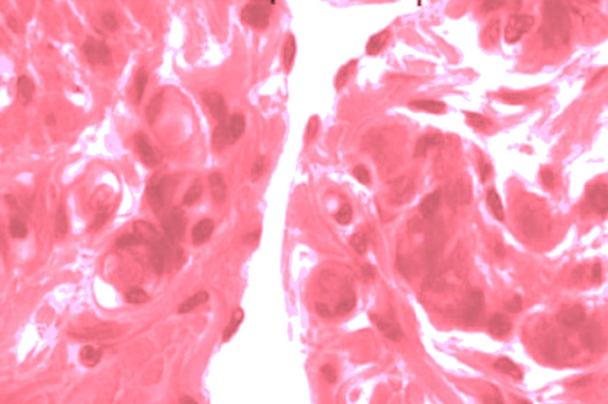
In the study of injected histological preparations, the most significant changes in the microvascular bed were detected in those regions of the intestinal wall with shared blood supply. Moreover, these areas are located mesoperitoneally and are therefore functionally inactive. In the microvascular bed of the submucosal base of these regions, signs of impaired vascular permeability were identified (Fig.3), with a shift of the gradient toward both arterial and venous microvessels. Loss of blood plasma was accompanied by hemoconcentration changes. However, the formation of thrombi in venules was not observed. Along the length of numerous arterioles, a sharp reduction in diameter was recorded, indicating cessation of bleeding within them. The diameter of venules, on the other hand, was reduced in a fragmentary manner, characterized by alternating constricted zones resembling peristaltic waves (Fig.4). Extravasation in the microvessels of the muscularis was less pronounced, and bleeding within them remained minimal throughout the experiment. Extravascular and rheological changes, as well as alterations in blood flow within the microvessels of the muscularis, were also less evident during the study. The superior mesenteric artery differs from other major arteries by the range of its vascularization. With impaired perfusion of the intestinal wall’s microcirculatory bed, the velocity of blood flow in the superior mesenteric artery decreases, and the diameter of its main trunk becomes reduced.
Data characterizing the microcirculatory bed of the greater omentum indicate that a significant volume of blood plasma leaks beyond its boundaries into the peritoneal cavity. The structural organization of these vessels resembles the glomerular capillary network of the renal corpuscle (Fig. 5). Thus, the results of experimental investigation into the morphological structures of blood circulation in the large intestine demonstrate that, following blood withdrawal, angiograms reveal a reduction in the diameter of the main trunk of the superior mesenteric artery and an increase in the duration of the arterial phase. In the study of injected histological preparations, the most significant changes in the microvascular bed were detected in those regions of the intestinal wall with shared blood supply. This provides grounds to consider the greater omentum as a donor of peritoneal fluid or even as the “third kidney” of mammals. In the microvascular bed of the submucosal base of these regions, signs of impaired vascular permeability were identified. Loss of blood plasma was accompanied by hemoconcentration changes. However, the formation of thrombi in venules was not observed. Along the length of numerous arterioles, a sharp reduction in diameter was recorded, indicating cessation of bleeding within them. The diameter of venules, on the other hand, was reduced in a fragmentary manner, characterized by alternating constricted zones resembling peristaltic waves.
CONCLUSİON The results of this study demonstrate that morphometric analysis of the vascular system of the large intestine is of great importance for evaluating its functional state both under normal conditions and in experimental settings. Normative parameters reveal the distributional characteristics of vessels in different segments of the intestine, while experimental models allow the observation of morphological changes in vessels during hemorrhage and perfusion disturbances.
The findings obtained:
- Expand knowledge on the pathogenesis of ischemic and post-ischemic changes [7].
- Provide opportunities for improving diagnostic and therapeutic strategies in clinical gastroenterology [8].
- Indicate that monitoring morphometric parameters of the microcirculatory bed may serve as a reliable marker for early detection of perfusion disorders [9,10].
- Show that this approach can be integrated into both experimental translational research and clinical practice through the application of endoscopic and angiological methods [11,12,13].
Thus, morphometric analysis of the vascular system of the large intestine is distinguished not only by its theoretical relevance but also by its practical applicability in clinical gastroenterology, providing a reliable foundation for future translational research.
Фигуры
Ключевые слова
Ссылки
1. Golperin YuM, Lazarev PI. Digestion and homeostasis. Moscow: Ripol Klassik; 2020. 303 p.
2. Granger DN, Holm L, Kvietys P. The gastrointestinal circulation: physiology and pathophysiology. Compr Physiol. 2015;5(3):1541–83.
3. Xu H, Mao C, Liu J, et al. Microanatomic study of the vascular and duct system of the submandibular gland. J Oral Maxillofac Surg. 2011;69(4):1103–7.
4. Directive 2010/63/EU of the European Parliament and of the Council on the protection of animals used for scientific purposes. St. Petersburg; 2012. 48 p.
5. Markov II, Petrov ES, Markova VI. Universal method for selective identification of argyrophilic structures. Morphological Bulletin. 2016;(1):114–7.
6. Babayeva RE. Morphological features of neurons innervating different parts of the large intestine. World Med Biol. 2021;(2):163–7. doi:10.26724/2079-8334-2021-2-76-163-167.
7. Keskin N, Bamac B, Cakir O, Colak T, Ors A, Barut C. Morphometric analysis of the inferior mesenteric artery using magnetic resonance angiography. Bratisl Med J. 2025;126(12):2107–15.
8. Gastrointestinal Endoscopy. Morphometric analysis of the blood supply to the stalk of pedunculated colon polyps. Gastrointest Endosc. 2013;77(3):455–62.
9. Slavu I, Filipoiu F, Tulin R, et al. Arterial vascularization of the colon: a guide to surgical resection. J Mind Med Sci. 2024;11(2):145–53.
10. Mareev GO, Mareev OV, Kapralov SV, Klimenko GA, Kalyuta TY. Laser Doppler flowmetry in the diagnosis of intestinal ischemia severity. Russ Open Med J. 2025;14(1):e0101.
11. Dickson K, Malitan H, Lehmann C. Imaging of the intestinal microcirculation during acute and chronic inflammation. Biology (Basel). 2020;9(12):418.
12.Haas G, Fan S, Ghadimi M, et al. Different forms of tumor vascularization and their clinical implications: vessel co-option in colorectal cancer liver metastases. Front Cell Dev Biol. 2021;9:612774.
13.Ortenzi M, Carsetti A, Picchetto A, et al. Implementation of intraoperative procedures to prevent anastomotic leak in low anterior rectal resections: novel technique to evaluate anastomotic vascularization. Surg Endosc. 2025;39(4):1123–31.
О статье:

История публикации

Опубликовано: 13.Oct.2026

Авторское право
© 2022-2025. Azerbaijan Medical University. E-Journal is published by "Uptodate in Medicine" health sciences publishing. All rights reserved.Связанные статьи
Просмотрено: 14